
Pyometra- Accidental finding on ultrasound six weeks from mating
Here you see a scan image of Pyometra, it was an Accidental finding on ultrasound 6 weeks from mating, The scan was preformed to check for pregnancy but in this
Here you see a scan image of Pyometra, it was an Accidental finding on ultrasound 6 weeks from mating, The scan was preformed to check for pregnancy but in this

Here you can see a sac that has been captured absorbing at day 34 of pregnancy, as you can see it looks the shape of a Doughnut, after a couple

Clear dog scan at 34 days gestation using the SIUI APOGEE 1000 Notice how clear the sacs can be seen on the video, Also i will include a still image


F: Left ventricular wall thickness measures within normal limits (septal wall thickness > posterior wall, which is common in cats). Good systolic function. Type 1 diastolic filling pattern. Trivial mitral
D (first video): Normal size left ventricle and wall thickness. Good biventricular function. No valvular stenosis or significant regurgitation. E (second video): Normal size left ventricle and wall thickness. Good

Normal size left ventricle and wall thickness. Good systolic function. Ejection fraction visually estimated at 70%. Normal diastolic function. Normal size left atrium. Normal size right atrium. Normal size right

Normal size left ventricle with increased wall thickness (6.1mm). Please note this has not been adjusted for weight Decreased septal and lateral E’ velocities (below 5cm/s), suggestive of diastolic dysfunction

A scan by Dr Simeon Konsulov, mobile veterinarian in Crewe. This cat is approximately 6 weeks pregnant.

A recent dog pregnancy scan by our experienced veterinarian, Simeon, of Mobile Vets Crewe.


This dog travelled from St Mary Cray for a scan today. We saw at least 7 puppies (almost certainly a lot more!), all beautiful and healthy at this stage. The
Beautiful images today performing a dog pregnancy scan. Saw 4-5 healthy puppies (could be more hiding!), all with strong heartbeats.
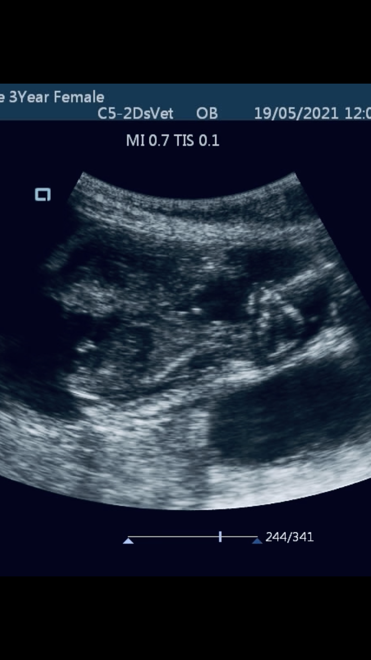
During this canine pregnancy scan (approx. 6 weeks post-mating) I found fluid inside the uterus, but no pregnancy. This should be checked by a veterinarian, as may need antibiotics to
I obtained a short axis view of the aortic valve first with a linear probe, due to the fact that I was scanning a tiny animal and wanted the highest
Another incredible images of a Labrador embryo – gestation age 6 weeks 2 days.
Amazing image of a Golden Doodle, gestation age, 6 weeks. Placenta is nearly as big as the pup!
Abdominal scan on a French Bulldog from Croydon, identifying bladder, intestines, liver and kidney.

Bella came all the way from Clapham today for her pregnancy scan. She is around 5 weeks pregnant and has absolutely beautiful images! We identified at least four puppies, all
