Alpaca Pregnancy Ultrasound Scans
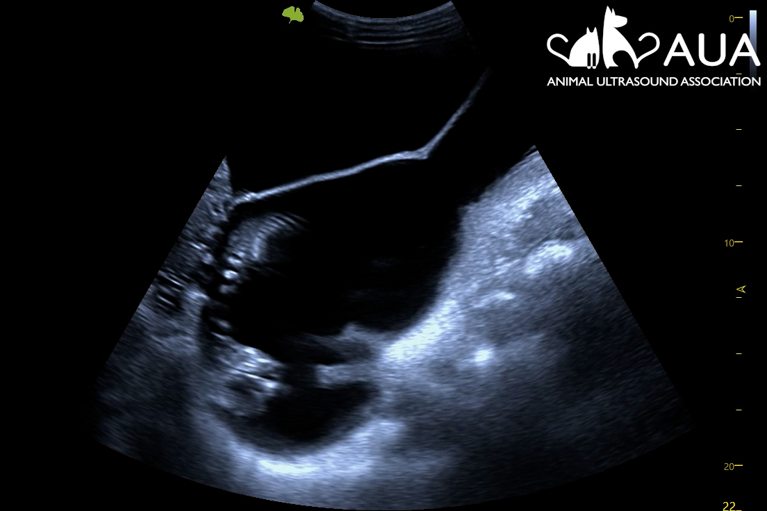
We confirmed 8 pregnancies today in Maidstone, Kent. Most of them were between 50 – 70 days, and it is surprising how tiny the foetus still is even at that


We confirmed 8 pregnancies today in Maidstone, Kent. Most of them were between 50 – 70 days, and it is surprising how tiny the foetus still is even at that

Dolly attended ten days ago for a 25-day scan. 25 days is too early to confirm pregnancy for an owner, but in Dolly’s case, she was scanned on the understanding

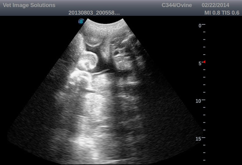
Nala came for her pregnancy scan today. We saw at least 6 (probably 7) foetuses, at around six weeks of gestation.