Ultrasound is a sound wave, and all sound waves are longitudinal waves. The way that sound energy travels through the body is akin to energy traveling along a slinky; if you hold it at one end and push, you will see a wave travel through it. This is the type of wave coming out of your ultrasound transducer – a wave that has a series of compressions and rarefactions. These waves travel out of the transducer in a fan shape. The sound waves travel from the probe, through your bitch to the pregnancy, and back again. It is bounced back, just like an echo.
A sine wave is often used to demonstrate aspects of sound waves, such as frequency and amplitude, but it’s important to be clear that sound is not in itself sine wave.
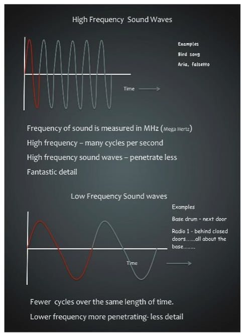
Watch Yvette use a sine wave to explain the concept of frequency below:
Coloured in red is one wavelength, or one cycle, with time along the bottom.
Transducers
Below is a diagram of the probe, and this is the most valuable part of your scan machine. The reason this is so valuable is the inside; inside this little head is a row of ceramic crystals. They have a very special job in that when electricity is applied to them, they vibrate. That vibration causes a sound wave to come out of the probe and into the body through a couplant. Between the scan head and the soft tissue, we have a couplant which is your ultrasound gel, and without this the ultrasound would just bounce back.

Our ultrasound wave comes out of here because the crystals have vibrated and produced it. It goes along it, hits the surface and then it bounces back to the probe and it hits those crystals again. The crystals vibrate again, and this time instead of turning an electrical signal into an ultrasound wave, they’re turning an ultrasound wave into an electrical signal. That is coming back to your scan machine and being displayed on here as a black and white image.
The kind of reflection that you get on your scan machine depends entirely on the surface that the ultrasound wave has been reflected from. Imagine a bucket of water and a pile of bones. If I put my ultrasound into the bucket of water there’s nothing much in the water to stop it continuing down to the bottom of the bucket, so it won’t bounce much back to my probe until it hits the bottom of the bucket. With the pile of bones, every time an ultrasound wave hits a bone it will bounce back to my screen. What I see then is a bright white line. So anything that’s bright is a good reflector which means it is solid. Anything that is black doesn’t return any echoes so the absence of reflections on our screen is black. So when you look at your bitch’s bladder and it’s full of pee there’s nothing in the bladder to hit and bounce back the sound until it hits the bladder wall – the bottom of the bucket.