
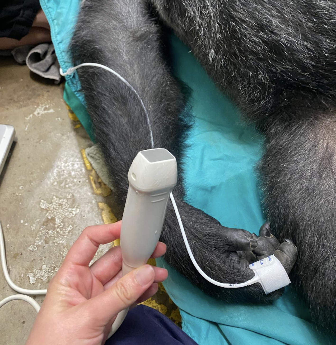
I was lucky enough to be invited to perform an echocardiogram on a gorilla over the summer, almost a year on from the very first gorilla I scanned.
The silverback I scanned in 2025 was 177kg at the time, which made for a very challenging echocardiogram. I had done my research, read the protocol from London Zoo and The Great Ape Project, memorised the probe positions, but my images were… not great. I knew his sheer size was the major factor, but nevertheless, I was disappointed; opportunities like this do not come around often and I was scared I might not get another.
I was therefore delighted, but nervous, when I was recently invited back to scan another gorilla. My expectations of getting decent images this time around were low, but I prepared as best I could, including lifting and holding heavier weights in the gym to improve my arm and shoulder strength and stamina! If you perform echocardiograms, you will understand my logic – it’s not because we press so hard into our patients; rather, it’s the holding of the probe for prolonged periods in one position that results in fatigue, particularly when the position is an unfamiliar one. When veterinarians attend our in-person echocardiography courses, it’s easy to see which views are new to people and which ones they practice regularly. Usually, it’s the apical views that people perform much less frequently, and subsequently their arms begin to fatigue quite quickly when practicing these views.
I am well used to holding the usual probe positions in cats or dogs, but I am out of practice on great apes, having not performed regular human echocardiography for many years – so gym sessions and practicing on my husband (who luckily did not take offence at being my substitute gorilla) was the best I could do.
With my image quality expectations sufficiently low, I travelled with my ScanX Vinno D10 and the lowest frequency probe I have, to scan my second ever gorilla.
This time though, I was scanning a female. She is almost 100kg lighter than her mate – and what a difference this made!
Above: Compared to the images I’d acquired a year earlier on a male silverback, my images this time around were a lot clearer
Above: Confidentially understandably surrounds these opportunities, but the first gorilla I scanned made it into the New York Post and BBC News – no longer such a secret!
Neither of the animals I scanned had known heart disease, so why perform an echo?
Captive gorillas are highly susceptible to heart disease and it is the leading killer of captive male gorillas. The main aim of these opportunistic scans is to pick up early signs of left ventricular remodelling, fibrosis, or aortic disease. Without an echocardiogram, the first sign of heart disease can often be sudden death.
Sedating gorillas is a huge production, and a masterclass in teamwork. It involves months of planning and training. In this situation, the gorilla had been trained to allow the keeper to administer the anaesthetic, but contingency plans always have to be in place in case she were to change her mind on the day. The other factor in the equation was her infant – could the baby be tempted away from her mother to play in the gym, or would she be distressed and need to be kept in an area beside her mother during the scan?
Luckily for us, all of the anticipated hiccups were averted: the gorilla willingly offered her arm to the keeper, and her infant was happy to go and play in the gym with her family, but the silverback of the group took an unexpected interest in proceedings and did not want to leave the side of his partner and there was a brief moment of tension in trying to coax him away!
Gorilla hearts are more trabeculated than human hearts, particularly at the apex, and there’s a fantastic paper from Aimee Drane on this. She shows that there is a negative (curvilinear) relationship between degree of trabeculation of the left ventricle, and systolic twist, such that the human heart has a more pronounced “wringing” motion during systole than other great ape hearts.
A healthy gorilla heart, looked at through human eyes, could be mistaken for a diseased one: the apex is so heavily trabeculated that some normal gorillas exceed the human threshold for left ventricular non-compaction. Whether we are scanning a gorilla, a greyhound or a Maine Coon, an echocardiogram is only as good as the reference values we interpret it against. Just as with our feline HCM patients, where the first sign of disease can be sudden death, a baseline scan in an apparently healthy animal is often the most valuable one we will ever do.
© 2026 | AUA – All Rights Reserved
