The umbilical veins carry oxygenated blood from the placenta to the foetus, and the umbilical arteries carry deoxygenated blood from the foetus back to the placenta. Dogs have two of each, unlike humans, who have only one umbilical vein.
Colour Doppler can tell you which vessels are the umbilical veins and which are the arteries by looking at the direction of blood flow. Flow moving towards the transducer will be coded in red, and flow away will be blue. Beginners often confuse this, incorrectly believing that blue blood flow on ultrasound is “deoxygenated” and red “oxygenated.” This is not the case, as you can change the colour of the observed blood flow simply by changing your transducer position.
Colour Doppler can be useful in differentiating structures which look similar on B-mode imaging – for example, the ureter or a vein – and can also be used by veterinarians to screen for pathology. Laminar flow, for example, is usually mostly all blue or all red (depending on direction), but turbulent flow will show as a mosaic of colours. Unexpected turbulence can alert the veterinarian to stenosis, or colour flow where none would be expected could suggest a shunt or anomalous connection.
Pulsed Wave Doppler, on the other hand, can be used to quantify flow. It allows us to measure the velocity at which the blood cells are travelling at different points in time.
Doppler does result in a higher TI (thermal index), so it is important not to use it during very early pregnancy when the foetal circulation is still immature.
The resistance index is a common measure used when examining pulsatile blood flow with Pulsed Wave Doppler (PW Doppler). It is calculated by measuring the peak systolic velocity, minusing the end diastolic velocity, and then dividing this all over the peak systolic velocity. A resistance index of 0 would be no resistance to flow at all, and 1 would be maximum resistance.
It is difficult to envisage what this means without giving it a go yourself. Practicing using PW Doppler on a large artery which is easy to find, such as your own carotid artery.
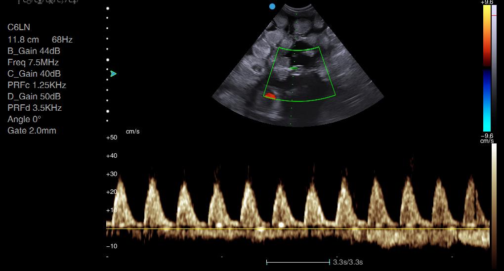
Step 1 : Find your carotid artery on B-mode. It’s easier with a linear probe, but the appearance will be similar with microconvex. You can tell this is pulsatile even before turning on colour Doppler.
Step 2: Turn on colour Doppler (the ‘C’ button on the Apogee 1000 Lite or Apogee 2300). There are two cine loops here – the first with the default colour map, the second after changing the colour map (C_Map on the menu) to one that I prefer.
By changing the angle of your transducer very slightly, you can remove some of the ambiguity in the colour coding. I’ve adjusted my angle so that most of the blood is flowing towards my transducer (albeit only just!), rather than perpendicular to it. This will be explained more fully in the colour Doppler webinar planned for early April 2021.
Step 3: Using colour to guide you, press ‘PW’ and position your pulsed wave Doppler gate over the vessel. Press ‘set’ or ‘PW’ again to activate. Use the ‘Base’ button to adjust your baseline so that all of your waveform is shown, and not cut off at the top or the bottom. If the velocity of flow exceeds the maximum velocity of your scale (i.e. your waveform does not fit within the area shown), you can press the ‘scale’ button to increase your scale.

You can identify the peak systolic and end diastolic velocities on your trace, though please note this is for practice/illustrative purposes only: absolute velocities are meaningless from this angle without angle adjustment.

If my peak systolic velocity is 40cm and my end diastolic is 10cm, then RI = 0.75.


Because we’re calculating an index, the units (cm/s or m/s) don’t matter, just so long as you’re consistent.
Giannico et al. (2015) calculated the resistance index in the umbilical artery in dogs. In a normal pregnancy, the resistance index steadily decreases throughout gestation. Giannico et al. (2015) looked specifically at the very end of pregnancy, to see whether changes in umbilical blood flow could be used to predict whelping date or birthing difficulties.
The authors found that the RI decreased to below 0.7 within the final 12 hours prior to delivery, dropping the most (to 0.56) between 6-1 hour before delivery.
Interestingly, where RI dropped but labour did not follow, the RI began to rise again after the time at which whelping should have occurred. The puppies also began to show signs of distress, as measured by M-mode of the foetal heart, with distress defined as a heart rate between 150-180 beats per minute.
“The lowest values of the RI in the umbilical artery occurred during the 48–24 h antepartum period. In other words, this would be the time that the bitch should have gone into labor, and when labor did not occur, there was a subsequent increase RI values above 0.7, until such time as the fetuses became distressed and were referred for cesarean delivery.”
Canine pregnancy scanning specialists are beginning to acquire more advanced machines, with high quality Doppler capabilities. Doppler studies of the umbilical arteries should not be performed as a commercial service outside of a veterinary practice, but may be of interest to breeders who use ultrasound to closely monitor their own bitches.
References
Giannico, A., Gil, E., Garcia, D., Froes, T. The use of Doppler evaluation of the canine umbilical artery in prediction of delivery time and fetal distress. Animal Reproduction Science, 154.