This was a tough video to film because my own dog’s heart is too confusing to demonstrate these views with (although she came in useful for the VSD part). We had to get our other office dog, who is a Golden Retriever, up onto the echo mattress, but the pulmonary valve can appear more distal in deeper-chested breeds. Don’t be confused if, when you try this view yourself, the pulmonary valve is not so low down on your screen as seen in my demonstration.
If you are not already subscribed to our echo newsletter, you can sign up here for free to receive new cases and learning resources approximately once a month.
A PDA is an abnormal connection between the aorta and the pulmonary artery, which in younger animals results in a continuous left to right shunt. You can hear this as a continuous murmur. As the disease progresses, the direction of the shunt can reverse, but this is extremely rare – the patient never usually makes it to this point.
The best views for seeing this are the right parasternal short axis view, optimised for the pulmonary artery, and the left-sided cranial position.
Ventricular septal defects are the most common type of congenital heart defects in cats, and a kitten with a loud murmur would immediately raise this suspicion.
A ventricular septal defect (VSD) is a gap (or defect) in the partition between left and right ventricles, called the “interventricular septum.” The result of this is that, when the ventricles contract during systole, the much higher systolic pressure generated by the left ventricle pushes blood out through the defect into the right ventricle, instead of it all going out through the aorta as it should.
This means that the heart has to work harder, but also that the normally low-pressure right ventricle, with this abnormal connection to the high pressure left ventricle, will increase in pressure over time. Surprisingly, though, as an isolated defect cats often tolerate a VSD surprisingly well and might never show any symptoms. Dogs can tolerate a small VSD well also, but more frequently have other abnormalities associated with it.
Due to their location – usually very close to the aortic valve – it is common for animals affected by VSDs to also have some degree of aortic regurgitation. In the demonstration video, I walk you through the views needed to identify a ventricular septal defect, as well as the other two most common defects detailed below. You will see that when I demonstrate a VSD on a Shetland Sheepdog, her aortic regurgitation has now become the dominant lesion.
An atrial septal defect (ASD) is a defect through the interatrial septum – the division between the right and left atrium. In a younger animal (unless the defect is huge) there will usually be a visible left to right shunt, but it will be of much lower velocity than a ventricular septal defect because the pressure gradient between the left and right atria is low.
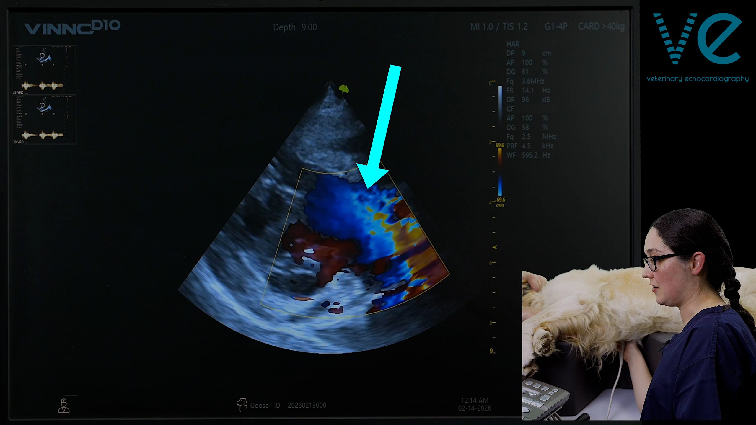
A patent foramen ovale (PFO) is also a gap in the interatrial septum but will be a lot smaller – often not always visible on 2D imaging – but you will still be able to detect the shunt by optimising your use of colour Doppler. The best method is to reduce your scale (PRF) to improve the sensitivity of your colour Doppler. Just remember to adjust it back afterwards!
Training small animal veterinarians in echocardiography is what I do. You can book a call with me, for free, to discuss what you’re stuck with, and we can figure out together how to get you to the next level and whether or not our training programme is right for you.
© 2026 | AUA – All Rights Reserved