
This week, we enjoyed another fantastic hands-on echocardiography workshop for veterinarians and RVNs in Bromley, led by Professor Luis Fuentes and Catherine Stowell. The main goals of the day were to improve confidence and reproducibility in image acquisition, and gain more practice with spectral Doppler.
We kicked off the day with a walkthrough on one of Virginia’s own dogs. Virginia pointed out the key landmarks of each view, before we broke into two groups based upon experience level.
Above: Virginia begins the day with a demonstration of each view and the landmarks she looks for.
At 11 o’clock, our first guest arrived: TikTok star Lyra, the Maine Coon. Lyra is a regular here and we know her to be normal, but she inspired a discussion about the diagnosis of hypertrophic cardiomyopathy in cats. Professor Luis Fuentes advised the group to look beyond solely wall thickness, and also look for other signs, such as obstruction and end-systolic cavity obliteration, particularly from the parasternal short-axis view. Recognising systolic anterior motion (SAM) was raised as a challenging skill, and this is covered extensively in the Confidence in Echocardiography training modules.
For end-stage HCM, Professor Luis Fuentes looks out for patchy, thinned walls and a fractional shortening below 30%.
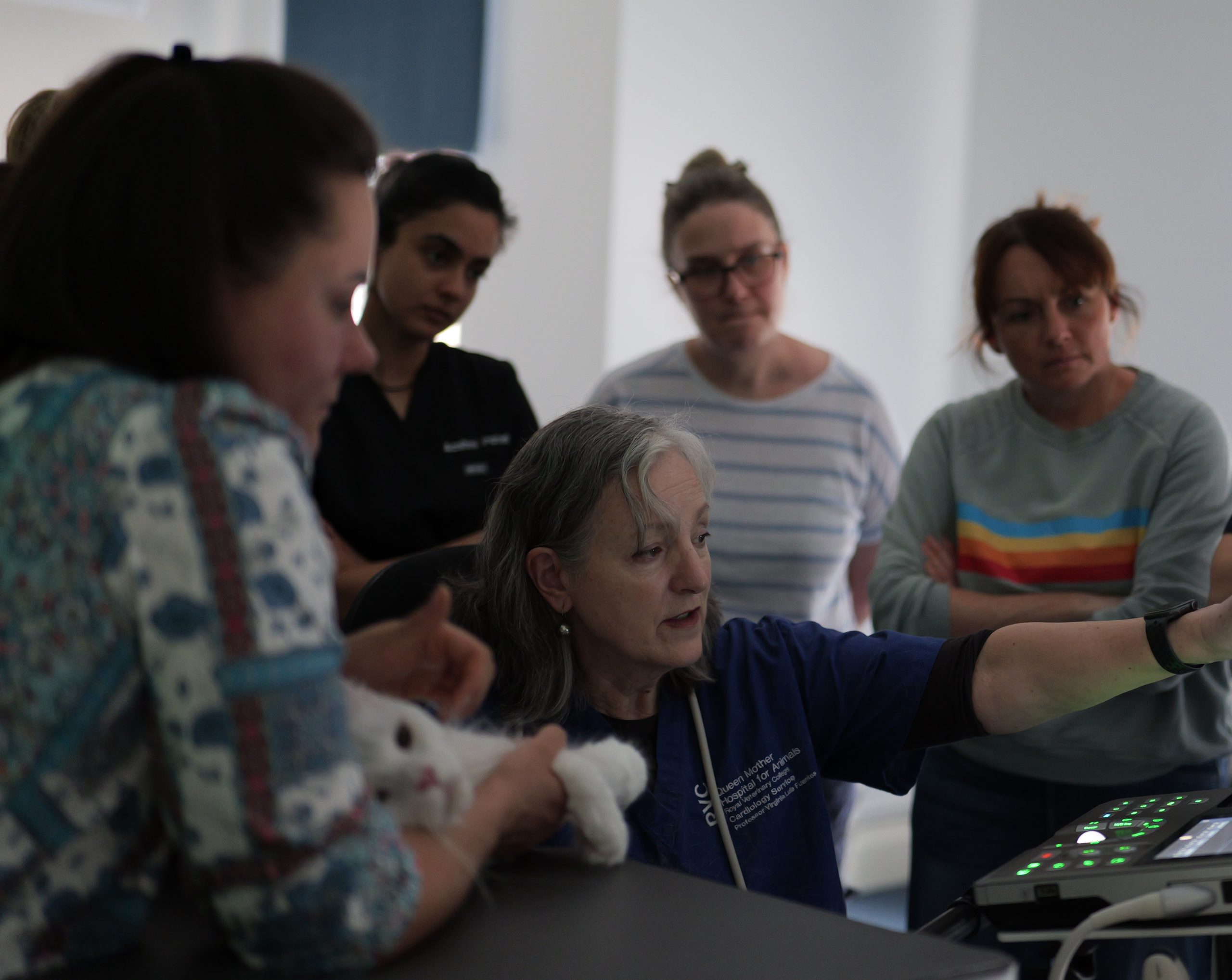
Above: Professor Luis Fuentes walks the group through how she performs a feline echocardiogram.
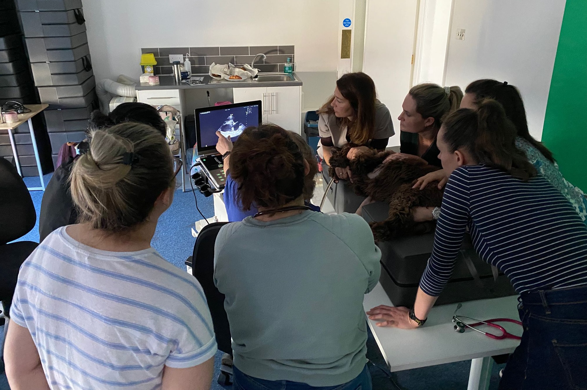
We were lucky enough to also have an 8 year old male Shih Tzu in attendance (with thanks to his owner and to Anderson Vets), giving us an opportunity to think about how our echo images could guide us in staging his myxomatous mitral valve disease. In our patient, cardiac remodelling had occurred, with a dilated left ventricle and left atrium. However, his E wave velocity on mitral inflow was only 0.9m/s, which is good news for him. If Virginia finds an E wave velocity of >1.2m/s (some people say 1.4m/s), she considers this patient to be at high risk of developing congestive heart failure in the near future.
Above: This Shih Tzu has significant mitral valve disease and cardiac remodelling (Stage B2).
This dog also did not appear to have a high probability of pulmonary hypertension at this stage. His right heart was normal in size with normal wall thickness, although we did find a tricuspid regurgitation velocity of just over 3m/s. If you are a Confidence in Echocardiography programme member, remember to register for our pulmonary hypertension webinar with cardiologist Dr Joshua Hannabus, being held at the end of the month. The below video might also be useful to you if you are still getting used to using continuous wave Doppler to examine the right heart.
After lunch, we had another visitor in the form of a young French Bulldog with unexplained seizures, whose echocardiogram turned out to be unremarkable. We also had a look at how to obtain a good view of the left atrial appendage, measure flow velocities with pulse wave Doppler, and discussed why this was important in cats with dilated left atria. If you are unsure about how to obtain this view, check out this video here:
If you would like to join the waiting list for our next echocardiography training day, we would love to hear from you!
If you would like to receive our monthly echocardiography newsletter, sign up for free here.
Ultrasound machines used: Vinno D10, Vinno D6, and Siemens P500.