

We are just coming off the high of a fantastic echo day with three amazing veterinarians, Professor Luis Fuentes, and some absolutely wonderful patients/volunteers. The combination of the hottest day of the year (at that point – I think that might have been beaten today!) and cats not liking the noise of the air conditioning unit had all the makings of a total disaster, but somehow we all survived it.
Our first case was a bit of a baptism of fire. Dollie first came to see me back in November, when her veterinarian heard a murmur at 12 weeks. Here was her original scan:
My summary report at the time read:
Increased left ventricular wall thickness, including apical hypertrophy.
Some degree of left ventricular outflow tract obstruction.
Right ventricular hypertrophy.
Right ventricular outflow tract obstruction, dynamic, mild-moderate.
Papillary muscle fibrosis, unusual appearance of mitral valve and apparatus, with regurgitation.
Prof Luis Fuentes reviewed the scan in December and agreed that the puppy had a hypertrophic cardiomyopathy phenotype and that the papillary muscle fibrosis was concerning, with a possible risk of sudden death. Without further tests, it was not possible to know whether this was genetic, or the result of a glycogen storage disease.
Dollie was re-homed locally (not sold) as the breeder wanted her close to keep an eye on her, and she attended 7 months later to our June echocardiography course.
Now 10-months old (and considerably more difficult to lift onto the echo table!), Dollie’s left ventricular hypertrophy has gone and the gradient through her pulmonary valve has reduced. She has a small amount of aortic regurgitation and Professor Luis Fuentes felt that her aortic annulus seemed a little small, but overall, a massive improvement from her first scan.
Virginia said it had been many years since she had seen such a case; some veterinarians would put these patients on atenolol and attribute the regression to the drug, but Virginia suspected that regression was, in fact, spontaneous in this exceptionally rare subset of patients – of which Dollie is one. Once the patient and her owners had left the room, the professor told us “you will probably never see a case like this again in your careers.” A good one to kick the day off with, then!
Next up were brothers Oliver and Harry. A local pet owner had been encouraged to bring them in for a scan after one of their relatives had recently been found to have a heart murmur. Although we have no further information on this heart murmur, with 1/7 cats having hypertrophic cardiomyopathy, this was clearly on our minds. Nevertheless, the expectation was that both young cats would be normal.
Have a look at some of the video clips some of our students obtained from Oliver, below. What do you think?
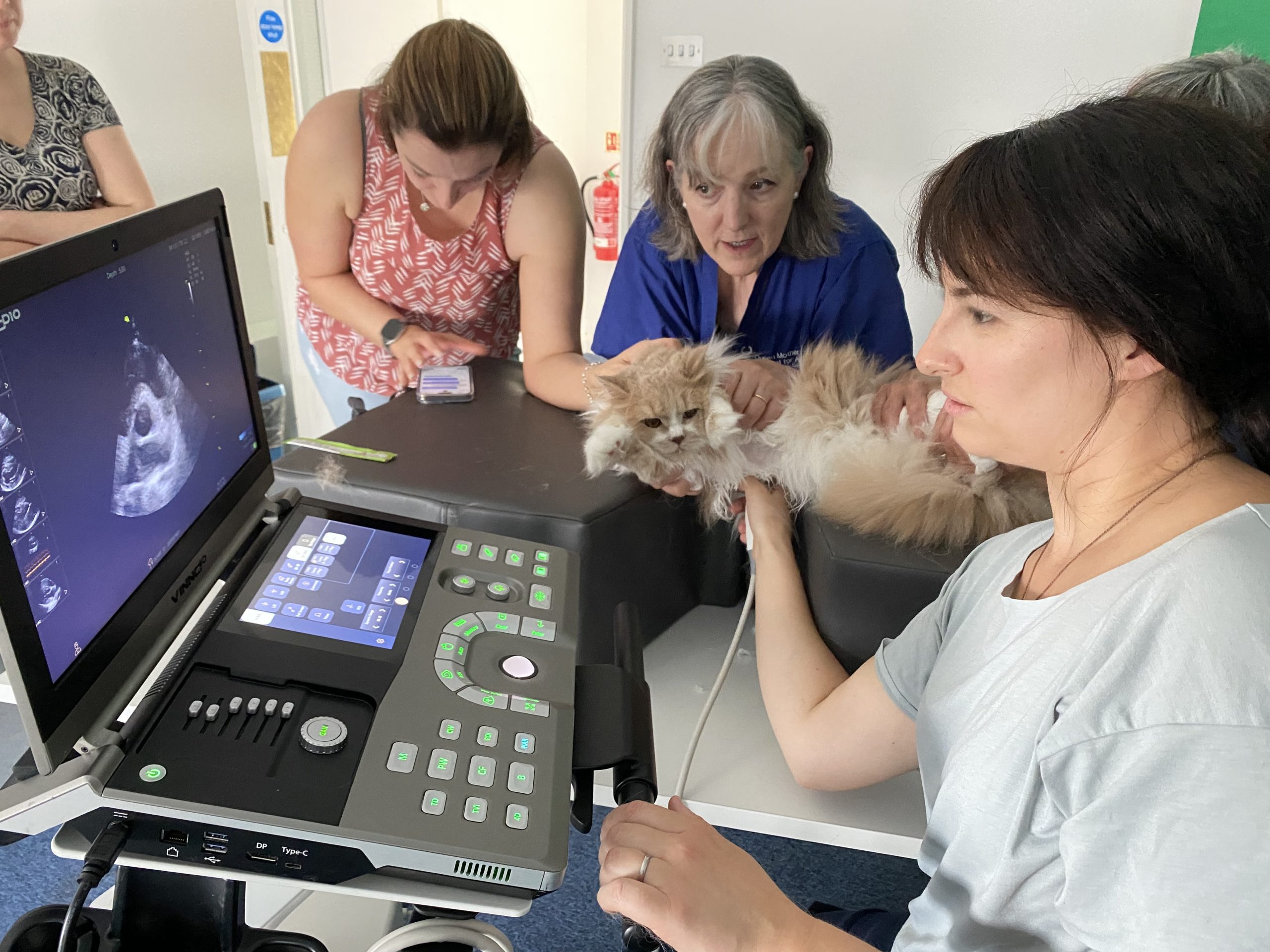
Above: Harry receiving his scan after Oliver.
What did you think of Oliver’s wall thickness? His left atrial size and function is normal, but his left ventricular cavity obliteration puts him in the “equivocal” category. While he might never progress, he is definitely one to keep an eye on, and the owner will bring him and brother Harry (who is currently totally normal) for regular checks in the future.
After the two British Longhairs, we had two sets of Maine Coons either side of lunch. Maine Coons are always so well-behaved for their echocardiograms, and two of them gave us extra practice time by falling asleep.
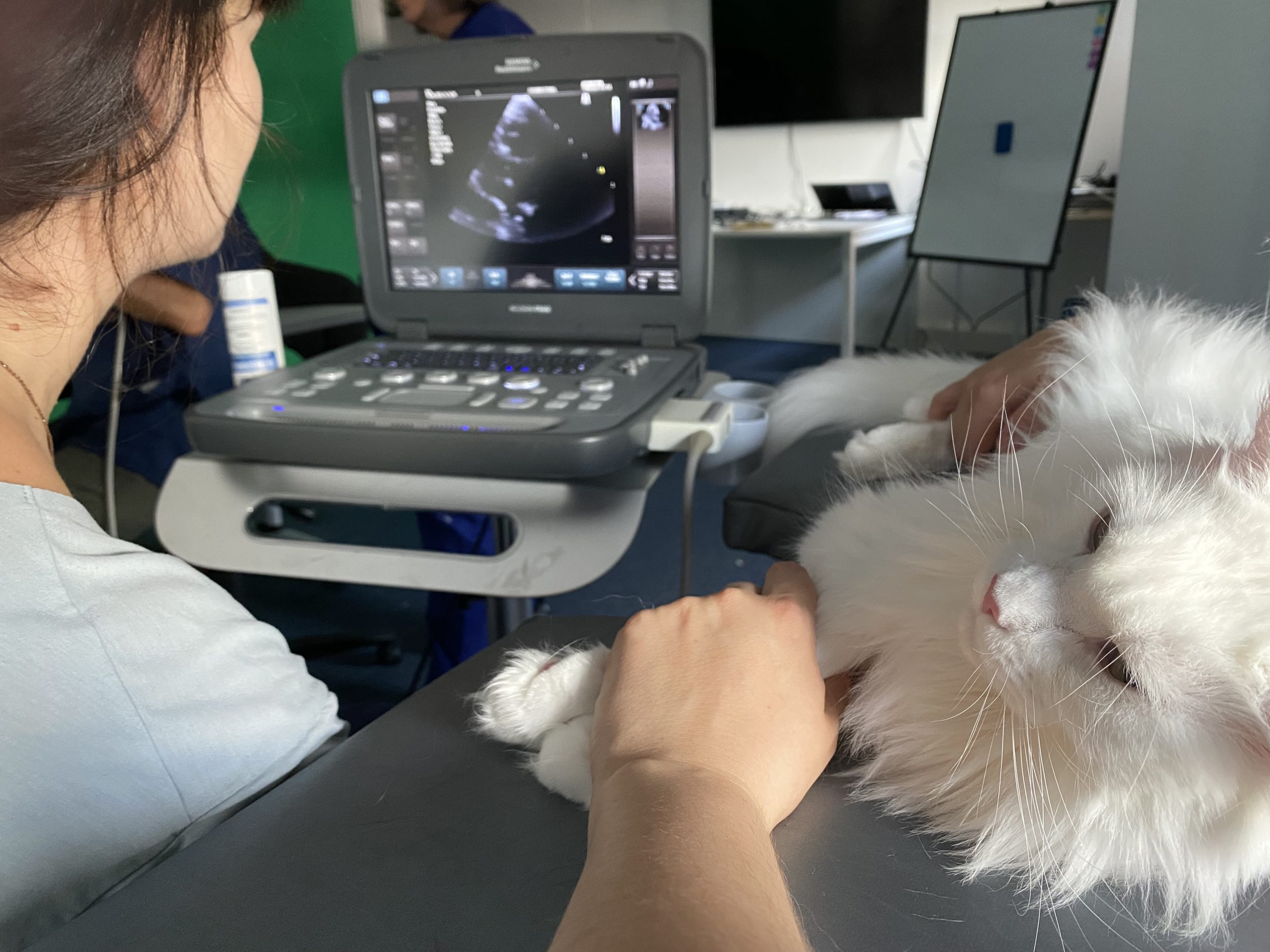
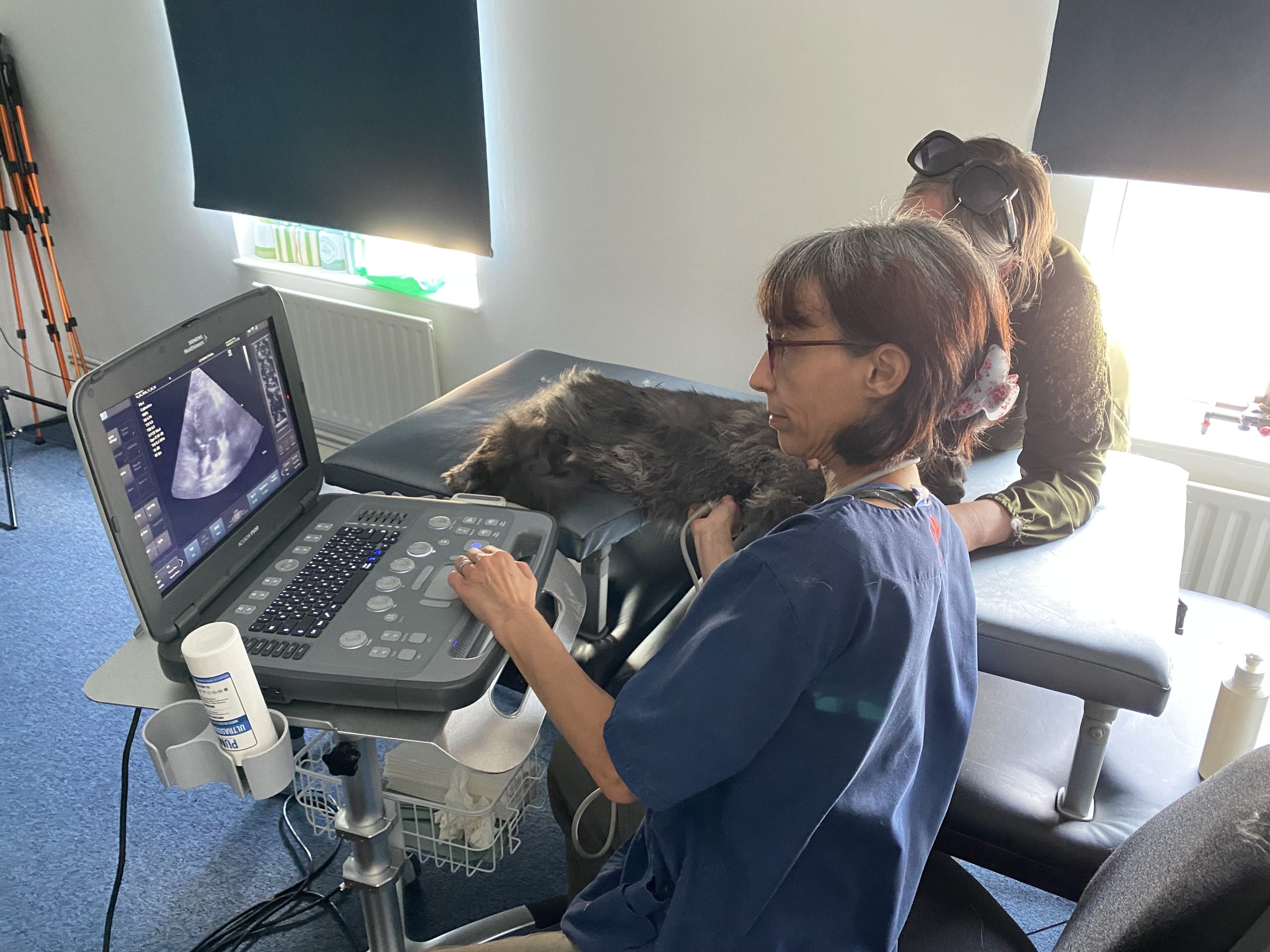
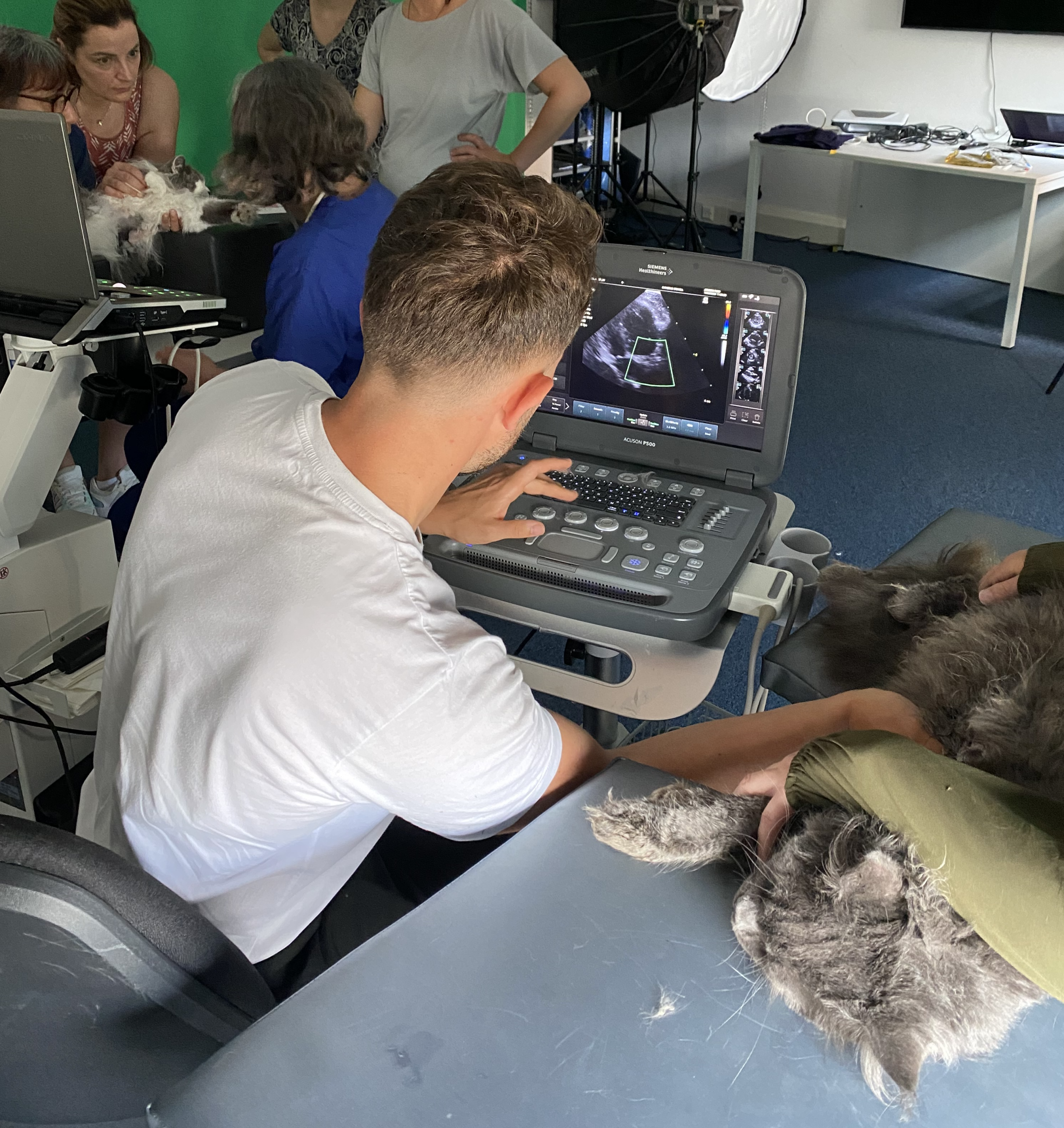
One of them was a follow-up scan for a previously discovered heart condition. Both this kitten and her brother had attended a previous echo course, and we found cardiac issues in both. Can you spot this kitten’s heart problem in the video below?
If you have some echocardiography experience then you probably noticed this kitten’s ventricular septal defect. There is some left ventricular dilatation, which is expected, but her heart otherwise looks fine and her prognosis is very good.
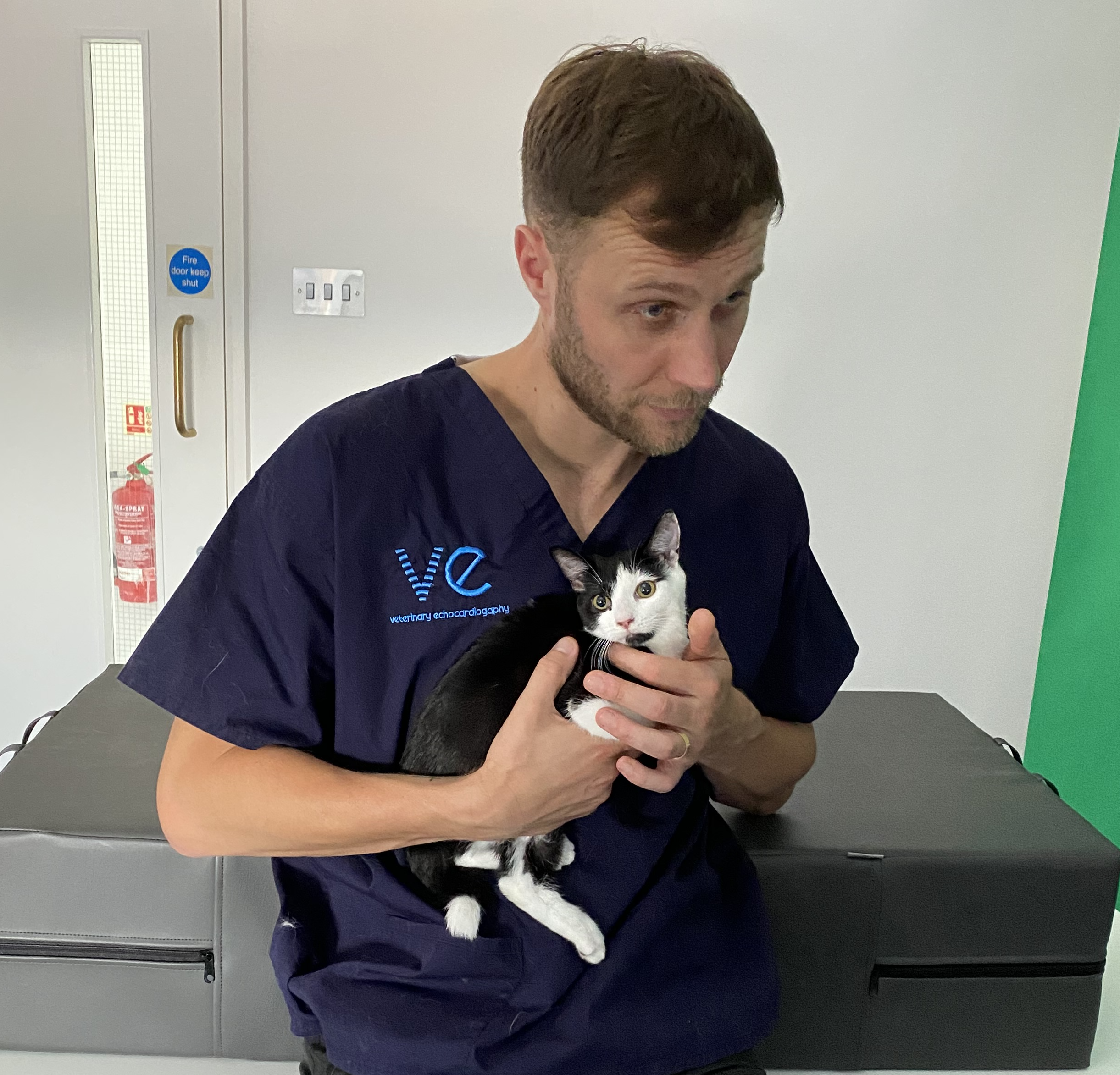
We also had Hercules in attendance, a tiny 6-month-old female kitten with a limb deformity, but thankfully a normal heart. She was so tiny, she had to be held to be scanned!
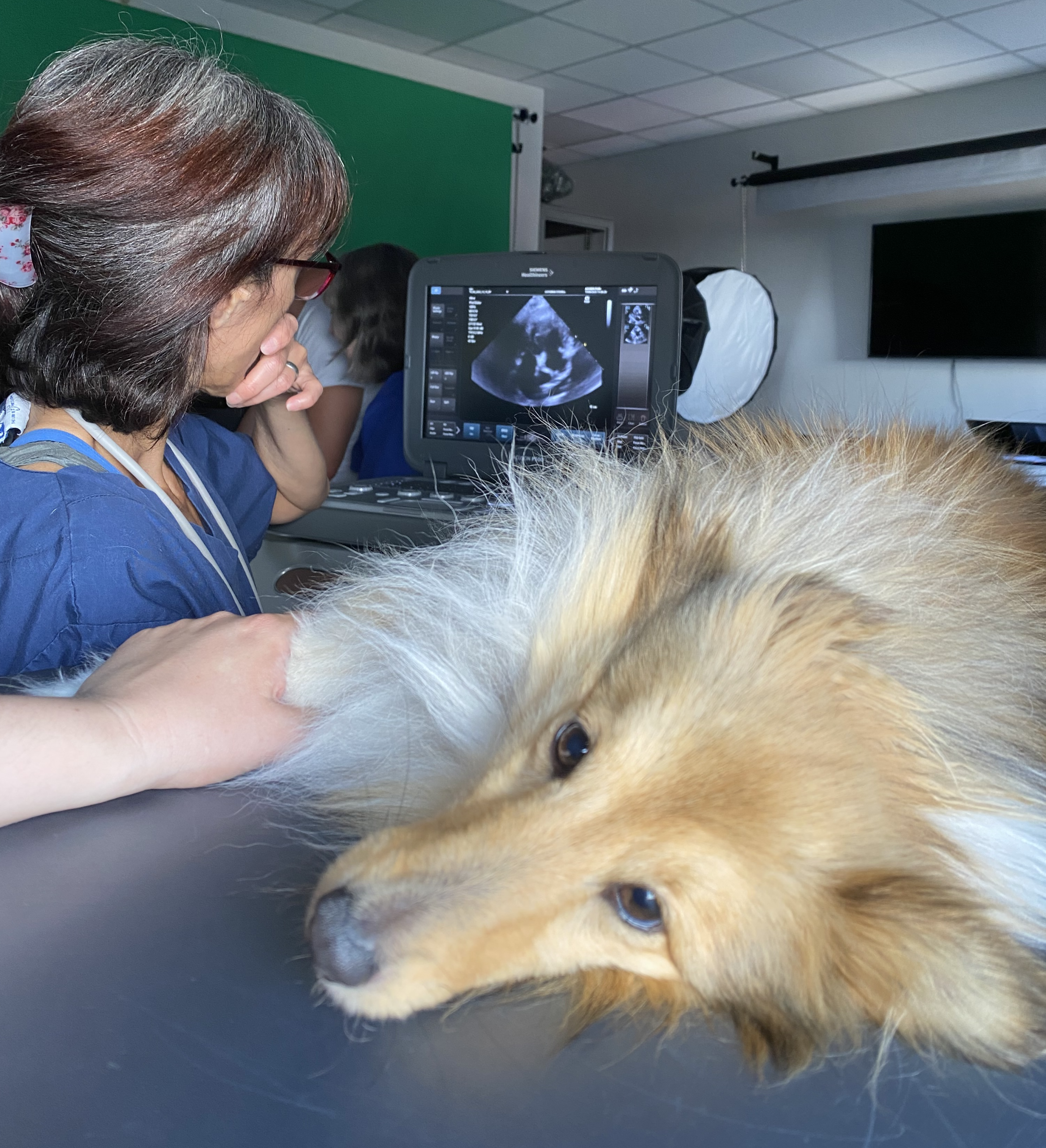
We finished off the day with two dogs – Virginia’s normal dog Bloss, and my very not normal dog and echo sidekick, Doppler.
Our hands-on echo days provide a unique opportunity to get precious practice time with animals in a calm, supportive environment. We make sure that at least 50% of the patients we see on the day are cats, because this is the species you are less likely to get the time with when you are back at work. We are exceptionally lucky that we are surrounded by supportive breeders and pet owners with very chilled out cats!
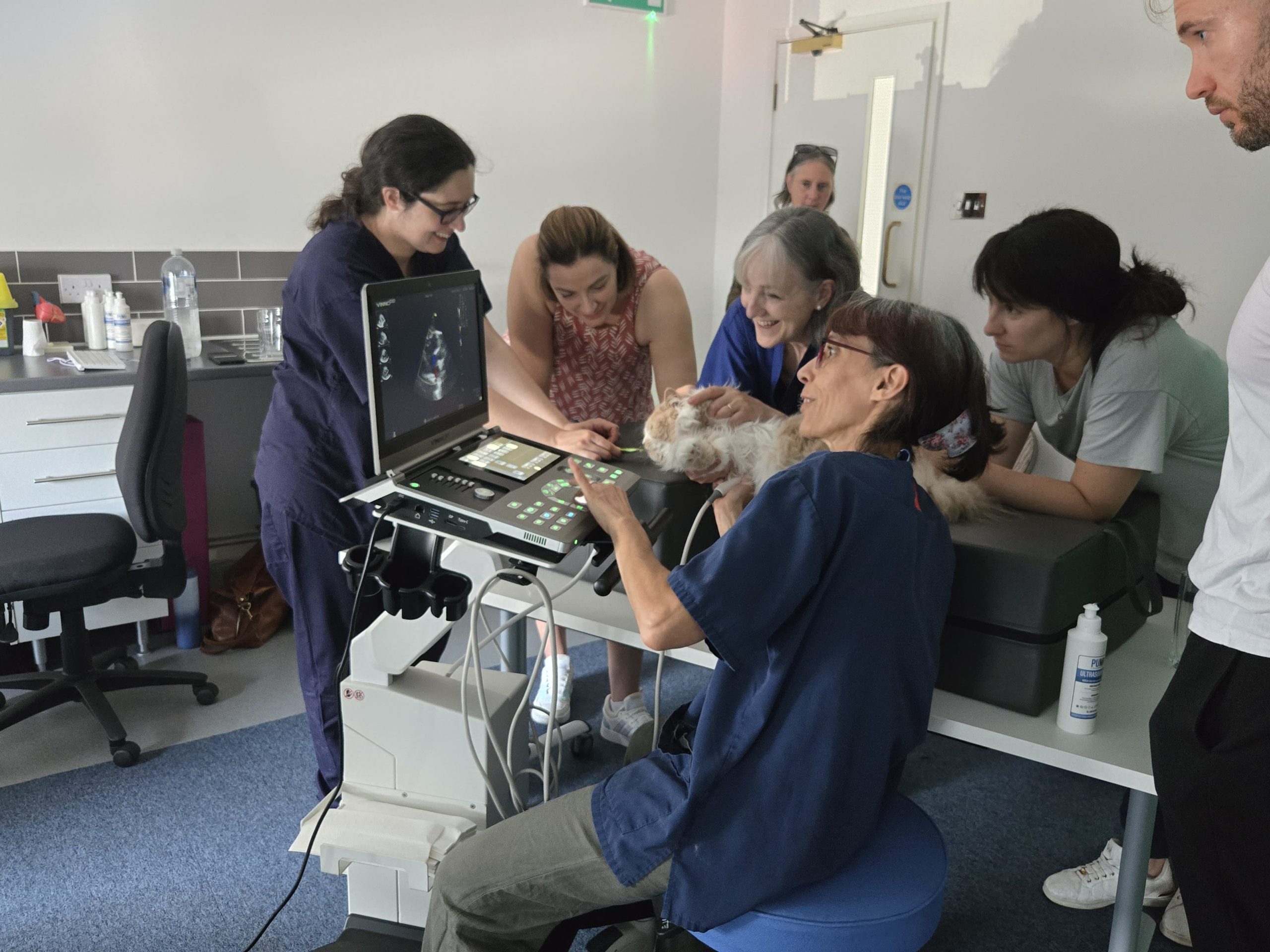
Nevertheless, a single day course is not a magic bullet. It needs to be followed up with structured learning, and supported with long-term guidance and mentoring, or everything you have learnt soon becomes a distant memory. It only takes a run of difficult patients to completely undermine your new-found confidence, and if you are learning echocardiography alone without expert guidance or the support of a community to help you learn from your images, it is very difficult to come back from those days.
If you would like to be kept up to date with our next in-person course dates, as well as receive case walkthroughs and teaching points directly to your email, you can sign up for our newsletter here. If you would like to find out more about how we can help you become confident with echocardiography, book a free Zoom call here.
Finally, for a full echo walkthrough showing all of the key views, you will hopefully enjoy this video below:
