Echocardiography Protocol Walkthrough for Scanning Dogs
Catherine Stowell
Share
All experienced sonographers follow a scanning protocol, whether their specialism is cardiac, abdominal, reproductive – it doesn’t matter. Not having this structure is one of the major factors that holds people back and slows their progress with ultrasound.
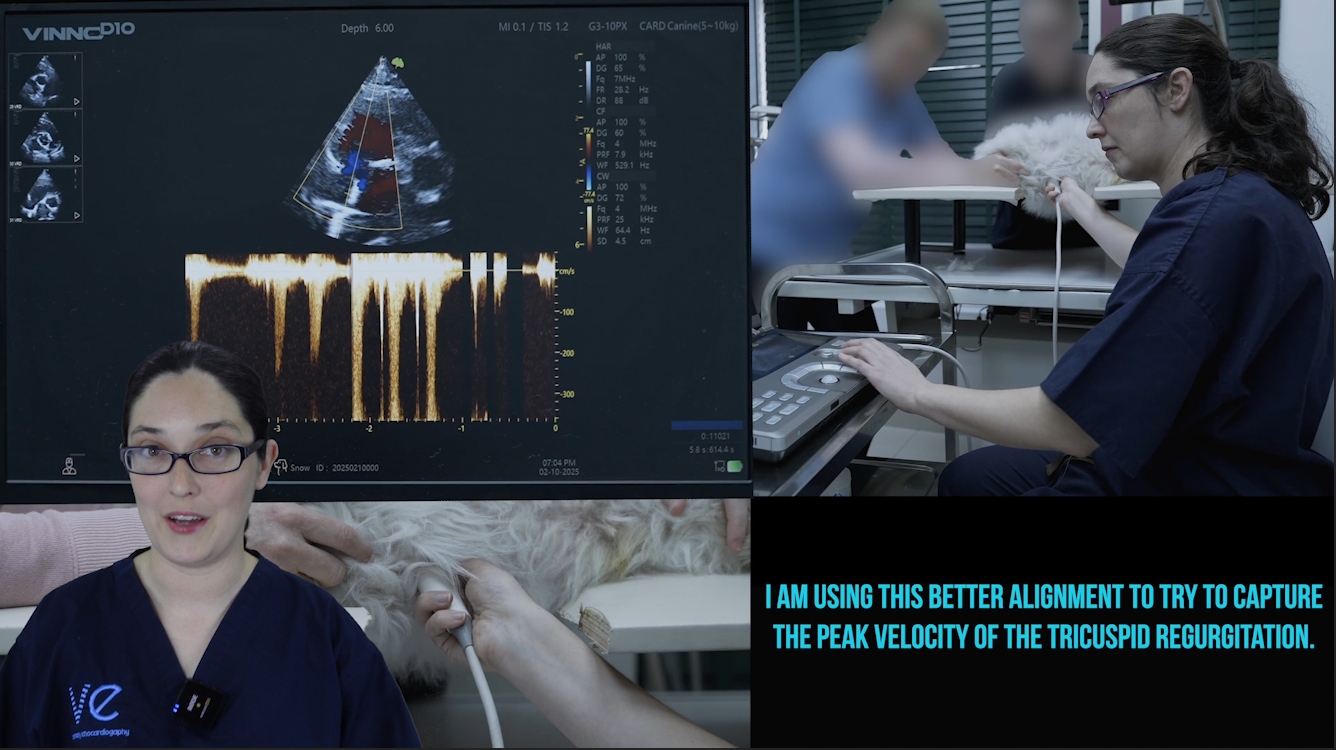
When you are performing an echocardiogram, you are already focusing on image optimisation, measurements, and you are definitely interpreting your findings as you go along. Not having to also think about what view comes next in your sequence is one less thing to worry about, and frees you up to focus on and improve the other parts of your examination. Of course, we frequently add in extra views, or modify standard ones, to investigate interesting findings more thoroughly – but we always have that framework to return to, to keep us on track.
The problem we have with veterinary echocardiography is that there is no set accreditation, guidelines or official protocol for cats and dogs, which can make it harder for people who are learning echo from scratch, or trying to progress to the next level. Teaching a set protocol is one of the things we focus on in the Confidence in Echocardiography coaching programme. I am always curious about how others perform their scans, so I thought it was about time I also shared ‘behind the scenes’ of how I go through a standard canine echocardiogram. You can watch the walkthrough here:
I skipped the subcostal view on this patient in order to keep the examination length down, given his breathing difficulties, but you can watch a demonstration of this here: